
Magnetic Speckle ImagingLensless imaging is on the forefront of technical
developments in synchrotron related studies. As the term suggests,
there is no optical system for imaging. Instead, a simple aperture
is used for limiting the illuminated area to the coherence length
of the beam (the light is usually in the longer wavelengths as the
lateral coherence length increases as a power of the wavelength). When the
illumination can be approximated by a plane wave, a detector
placed in the far-field measures a sort of "structure factor", which is
proportional to the square of the fourier amplitudes of the charge
(or magnetization) distribution within the area of interest.
The crucial step to obtain the real space image from this far-field
pattern is to retrieve the missing phases. The problem of phase retrieval
is common to all diffraction based structural studies (such as in x-ray
crystallography). Therefore, a lot of effort is devoted to fourier inversion
from incomplete data sets.
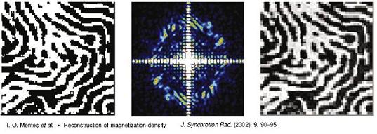 Figure 1: Shown in the middle panel is the simulated speckle pattern at the Fe L edge for a GdFe2 film with a magnetization distribution displayed on the left. On the rightmost panel, the result of the magnetic reconstruction from the simulated data reproduces well all the features of the original image.
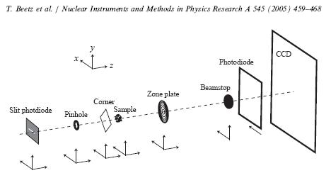 Figure 2: Sketch of the instrument. Note that the photodiode before the CCD detector is used as a diagnostic, and is removed during actual data acquisition. The beamstop is to protect the detector from direct x-ray beam. The pinhole is the actual beam defining aperture (about 10 micron diameter), and the zone plate is used only for direct imaging. The corner is a technical necessity in order to block out the diffraction from the pinhole aperture (i.e. the airy pattern from the circular pinhole).
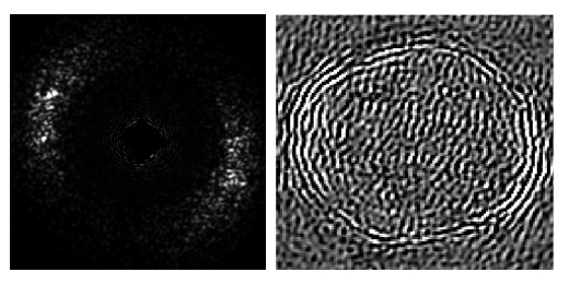 Figure 3: Magnetic speckles measured at the Advanced Photon Source ID-4C beamline. The left panel shows the magnetic contribution to the diffraction pattern of a Co/Pt multilayer sample illuminated with circularly polarized x-rays at the Co L3 edge (778 eV). The right panel is a real-space reconstruction of the out-of-plane magnetic domains [3].
[1] T. O. Menteş, C. Sanchez-Hanke, C. C. Kao, "Reconstruction of magnetization density in two-dimensional samples from soft X-ray speckle patterns using the multiple-wavelength anomalous diffraction method", J. Synch. Rad. 9 (2002) 90.
[2] T. Beetz, M. R. Howells, C. Jacobsen, C. C. Kao, J. Kirz, E. Lima, T. O. Menteş, H. Miao, C. Sanchez-Hanke, D. Sayre, D. Shapiro, "Apparatus for X-ray diffraction microscopy and tomography of cryo specimens", Nucl. Inst. Meth. in Phys. Res. A 545 (2005) 459.
[3] T. O. Menteş, "Imaging Magnetic Thin Films Using Resonant X-ray Scattering", Ph.D Thesis, SUNY at Stony Brook, November 2003.
|
