
A new milestone toward high-resolution clinical lung imaging
Imaging the smallest structures of the human lung remains a major challenge for medical imaging. While conventional computed tomography (CT) is essential for diagnosing and monitoring pulmonary diseases, its ability to reveal fine anatomical details is limited by the need to keep radiation exposure at clinically acceptable levels. Achieving substantially higher spatial resolution without a corresponding increase in radiation dose is therefore a key objective in the development of next-generation lung imaging. Synchrotron-based phase-contrast CT offers a promising approach to addressing this challenge, combining enhanced soft-tissue contrast with high-resolution three-dimensional imaging at radiation doses potentially compatible with clinical applications.
Researchers have now taken an important step toward this goal by successfully imaging a fresh, unfixed human lung obtained during forensic autopsy. The experiment was performed at the Synchrotron Radiation for Medical Physics (SYRMEP) beamline of Elettra Sincrotrone Trieste. Unlike conventional CT, which relies mainly on differences in X-ray absorption, phase-contrast imaging also exploits subtle changes in the X-ray wave as it passes through tissue, making fine structures easier to distinguish. Importantly, the imaging approach and experimental setup were developed with future clinical translation in mind, aiming to achieve substantially improved anatomical detail while maintaining radiation exposure within a clinically relevant range.
To perform the experiment, the researchers placed the lung in a specially designed human chest phantom, which reproduces key features of the thorax and keeps the organ gently inflated during scanning. The phantom was specifically developed to approximate important conditions of human chest imaging, providing a clinically oriented experimental environment for evaluating the technique. Combining this setup with the advanced X-ray imaging capabilities of SYRMEP, they obtained a three-dimensional dataset of the entire lung with an effective voxel size of just 67 micrometres — about the width of a fine human hair. Importantly, the estimated radiation exposure was comparable to that of conventional clinical CT, demonstrating that this level of anatomical detail does not necessarily require the substantially higher doses often associated with ultra-high-resolution imaging. This milestone extends earlier experiments with animal lungs to fresh human tissue without chemical fixation.
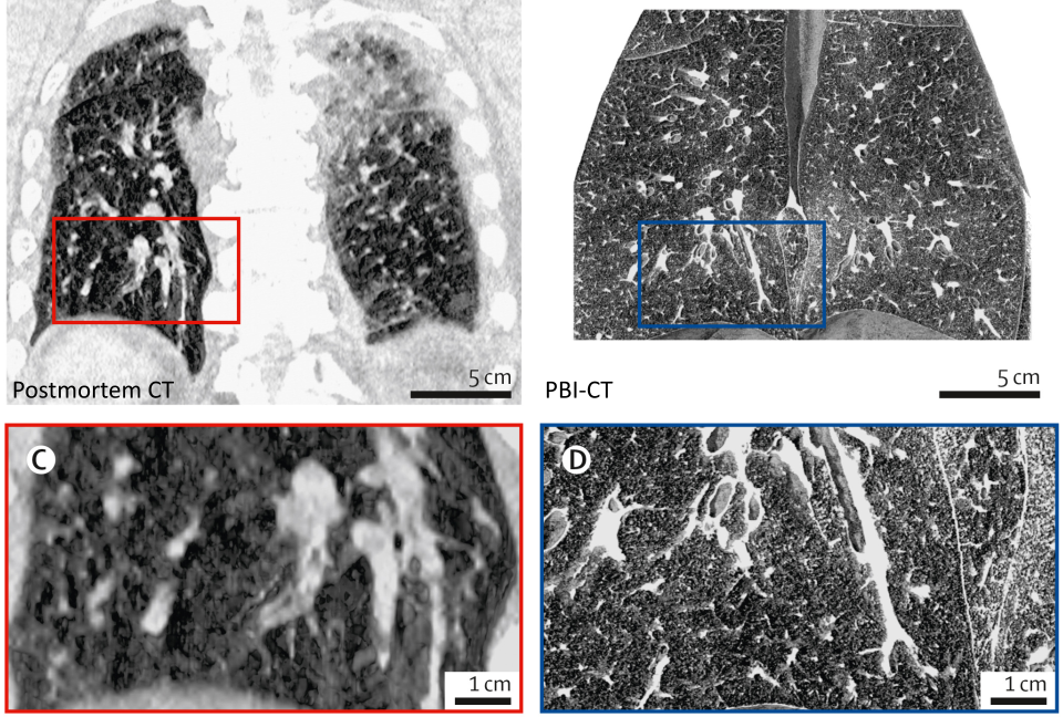
Figure 1: Comparison of conventional postmortem CT and synchrotron phase-contrast CT (PBI-CT) of the same human lung. The synchrotron images, acquired with the lung maintained under passive inflation, reveal substantially finer anatomical details, including small airways and pulmonary structures that are difficult to distinguish in conventional postmortem CT (CC BY 4.0 license).
The resulting images revealed small airways, fine blood vessels, and the delicate boundaries separating the lung’s structural units with remarkable clarity (Fig. 1). In a comparison with conventional postmortem CT of the same specimen, the synchrotron images (PBI-CT) showed substantially more anatomical detail and fewer extensive dense regions associated with the collapsed postmortem lung. Because the two scans were performed under different conditions — the conventional CT in the body and the synchrotron CT after removal and inflation of the lung — these differences reflect both the imaging approach and specimen preparation. The high-resolution, non-destructive images also made it possible to examine how tissue alterations varied throughout the organ, offering valuable information alongside conventional pathological examination.
Beyond forensic research, the study provides an important technological foundation for future lung imaging. The combination of high spatial resolution, enhanced soft-tissue contrast, and radiation exposure comparable to conventional clinical CT is particularly relevant for the potential translation of this technology to living patients. Visualizing delicate pulmonary structures throughout a whole organ could help researchers better understand lung disease and ultimately guide the development of more detailed diagnostic approaches. The work is also directly relevant to the planned SYRMEP–Life Science (SYRMEP-LS) beamline at Elettra, which is intended to support advanced lung imaging research and future proof-of-concept studies in selected patients. Although further development and clinical validation are needed, this demonstration of high-resolution whole-lung imaging under clinically oriented experimental conditions represents an important step toward the future application of synchrotron phase-contrast CT in patients.
The table-of-contents graphics shown on the homepage and in the top-stories archive represent an AI-graphically processed synchrotron radiation CT image rendered in false colors.
This research was conducted by the following research team:
Claudia Victoria Benke1,2, Christian Dullin1,2,3,4,5, Davide Radaelli6, Tommaso Bruscagin6, Deborah Bonazza6, Elena Longo3, Lorenzo D’Amico3,7, Jürgen Biederer1,2,8,9, Hans-Ulrich Kauczor1,2, Philipp Nolte10, Sam Bayat11, Giuliana Tromba3, and Stefano D’Errico6
1 Department of Diagnostic and Interventional Radiology, University Hospital Heidelberg, Heidelberg, Germany
2 Translational Lung Research Center Heidelberg, German Center for Lung Research (DZL), Heidelberg, Germany
3 Elettra-Sincrotrone Trieste, Basovizza, Italy
4 Translational Molecular Imaging, Max Planck Institute for Multidisciplinary Sciences, Göttingen, Germany
5 Department of Clinical and Interventional Radiology, University Medical Center Göttingen, Göttingen, Germany
6 Department of Medical, Surgical and Health Sciences, Hospital of Cattinara, University of Trieste, Trieste, Italy
7 European Molecular Biology Laboratory (EMBL), Hamburg Unit, German Synchrotron Research Centre (DESY), Hamburg, Germany
8 Faculty of Medicine, Kiel University, Kiel, Germany
9 Faculty of Medicine and Life Sciences, University of Latvia, Riga, Latvia
10 Faculty of Engineering and Health, University of Applied Sciences and Arts Göttingen, Göttingen, Germany
11 Inserm UA07 STROBE Laboratory, Université Grenoble Alpes, Grenoble, France
Reference
C. V. Benke, C. Dullin, D. Radaelli, et al., “Synchrotron phase-contrast CT of a human lung from forensic autopsy: a single-case technical feasibility study.”, The Lancet Medical Imaging & Theranostics (2026); DOI: 10.1016/j.lanmit.2026.100025.
